Home

סנקציה סליל לוח זמנים phospho histone h3 proliferation marker סט מודע אבזם

Research Techniques Made Simple: Techniques to Assess Cell Proliferation -  ScienceDirect
Research Techniques Made Simple: Techniques to Assess Cell Proliferation - ScienceDirect

Proliferation and Cell Cycle Dynamics in the Developing Stellate Ganglion |  Journal of Neuroscience
Proliferation and Cell Cycle Dynamics in the Developing Stellate Ganglion | Journal of Neuroscience

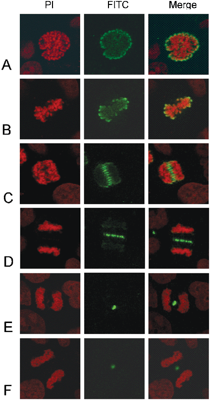
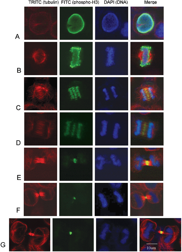
Dynamic distribution of Ser-10 phosphorylated histone H3 in cytoplasm of  MCF-7 and CHO cells during mitosis | Cell Research
Dynamic distribution of Ser-10 phosphorylated histone H3 in cytoplasm of MCF-7 and CHO cells during mitosis | Cell Research

Phosphorylation of histone H3 on Ser-10 in apoptotic cells. (A) and (B)...  | Download Scientific Diagram
Phosphorylation of histone H3 on Ser-10 in apoptotic cells. (A) and (B)... | Download Scientific Diagram

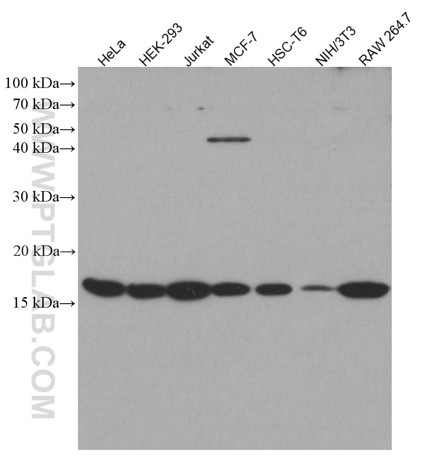
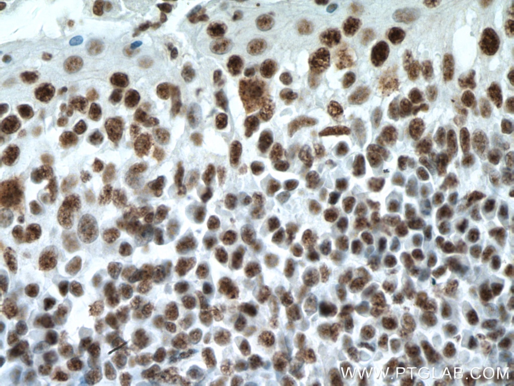
Phospho-Histone H3 (Ser10) Antibody 66863-1-Ig | Proteintech

Markers to assess proliferation and apoptosis. Identification of... |  Download Scientific Diagram
Markers to assess proliferation and apoptosis. Identification of... | Download Scientific Diagram

Phosphorylated Histone H3 (PHH3) Is a Superior Proliferation Marker for  Prognosis of Pancreatic Neuroendocrine Tumors | SpringerLink
Phosphorylated Histone H3 (PHH3) Is a Superior Proliferation Marker for Prognosis of Pancreatic Neuroendocrine Tumors | SpringerLink

Phospho-histone H3 (PHH3) staining shown some regenerating cardiac... |  Download Scientific Diagram
Phospho-histone H3 (PHH3) staining shown some regenerating cardiac... | Download Scientific Diagram

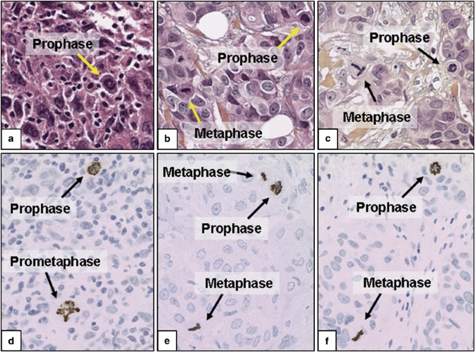
Phosphohistone H3 expression has much stronger prognostic value than  classical prognosticators in invasive lymph node-negative breast cancer  patients less than 55 years of age | Modern Pathology
Phosphohistone H3 expression has much stronger prognostic value than classical prognosticators in invasive lymph node-negative breast cancer patients less than 55 years of age | Modern Pathology
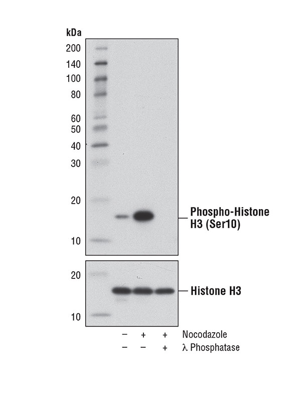
Anti Phospho Histone H3 | Cell Signaling Technology Inc | Bioz

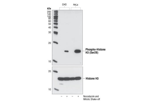
Phospho-Histone H3 (Ser28) Antibody | Cell Signaling Technology

Overview of Cell Proliferation | Cell Signaling Technology

Dynamic distribution of Ser-10 phosphorylated histone H3 in cytoplasm of  MCF-7 and CHO cells during mitosis | Cell Research
Dynamic distribution of Ser-10 phosphorylated histone H3 in cytoplasm of MCF-7 and CHO cells during mitosis | Cell Research

Ventricular zone cells express proliferation markers phosphorylated... |  Download Scientific Diagram
Ventricular zone cells express proliferation markers phosphorylated... | Download Scientific Diagram
Phospho-Histone H3 (Ser10) Antibody 66863-1-Ig | Proteintech

The proliferation markers Ki-67/MIB-1, phosphohistone H3, and ...

MicroRNA-7 Regulates the mTOR Pathway and Proliferation in Adult Pancreatic  β-Cells | Diabetes
MicroRNA-7 Regulates the mTOR Pathway and Proliferation in Adult Pancreatic β-Cells | Diabetes

Greater phospho-histone H3 staining after 56 Fe radiation. A)... | Download  Scientific Diagram
Greater phospho-histone H3 staining after 56 Fe radiation. A)... | Download Scientific Diagram

Phosphohistone H3 expression has much stronger prognostic value than  classical prognosticators in invasive lymph node-negative breast cancer  patients less than 55 years of age | Modern Pathology
Phosphohistone H3 expression has much stronger prognostic value than classical prognosticators in invasive lymph node-negative breast cancer patients less than 55 years of age | Modern Pathology

Dynamic distribution of Ser-10 phosphorylated histone H3 in cytoplasm of  MCF-7 and CHO cells during mitosis | Cell Research
Dynamic distribution of Ser-10 phosphorylated histone H3 in cytoplasm of MCF-7 and CHO cells during mitosis | Cell Research

Phospho-histone H3 (PHH3) labeling of mitotic cells. Sections examined... |  Download Scientific Diagram
Phospho-histone H3 (PHH3) labeling of mitotic cells. Sections examined... | Download Scientific Diagram

Double IF staining for PH3 (marker for proliferation) and E-cadherin... |  Download Scientific Diagram
Double IF staining for PH3 (marker for proliferation) and E-cadherin... | Download Scientific Diagram

Measurement of cell cycle entry activity by phospho-histone H3 (pH3)... |  Download Scientific Diagram
Measurement of cell cycle entry activity by phospho-histone H3 (pH3)... | Download Scientific Diagram

Proliferation marker phosphorylated histone H3 is decreased by rLOX-PP... |  Download Scientific Diagram
Proliferation marker phosphorylated histone H3 is decreased by rLOX-PP... | Download Scientific Diagram

Phosphorylated Histone H3 (PHH3) Is a Superior Proliferation Marker for  Prognosis of Pancreatic Neuroendocrine Tumors | SpringerLink
Phosphorylated Histone H3 (PHH3) Is a Superior Proliferation Marker for Prognosis of Pancreatic Neuroendocrine Tumors | SpringerLink

Proliferation marker phosphorylated histone H3 is decreased by rLOX-PP... |  Download Scientific Diagram
Proliferation marker phosphorylated histone H3 is decreased by rLOX-PP... | Download Scientific Diagram

Markers to assess proliferation and apoptosis. Identification of... |  Download Scientific Diagram
Markers to assess proliferation and apoptosis. Identification of... | Download Scientific Diagram